Medical school curricula: Anatomy Class is reinventing itself without cadavers
Anatomy class and dissection in particular are formative parts of medical education with a long history of the smell of formaldehyde and the emotional development around caring for your “first patient.” It is arguably a rite of passage. The use of cadavers in medical training dates back over 100 years. The idea of the cadaver as the first patient is explored in Body of Work: Meditations on Mortality from the Human Anatomy Lab by Christine Montross. Many schools have integrated funeral services or ceremonies to honor the bodies and integrate ideas around professionalism, empathy, and honoring of patients.
However, cadaver dissection has its controversies and challenges. Cadavers are often not a fantastic representation of a human, the textures and colors are different, and at times the normal anatomy is muddled by disease or decay. They are expensive and challenging to maintain and store. There is also controversy around how bodies are obtained for cadaveric dissection. Historically, cadavers were actually obtained by grave robbing poor, enslaved, or non-White bodies. In the essay, “An Unexpected Education-Unclaimed Bodies in the Anatomy Lab" published in the NEJM, Nov 19, 2020, Mary Peeler explores the ethical dilemmas around using “unclaimed” bodies in the anatomy lab. At the time of her essay writing, 12% of U.S. medical schools were still using unclaimed bodies as part of their anatomical classes and training. Unclaimed bodies are the remains of people lacking the ability to pay for burial costs. In New York City a measure was passed in 2016 that requires written consent from a spouse or next of kin before an unclaimed body is released to a school. This article in Academic Medicine looks at the sources of bodies for anatomy education worldwide. There is also a for-profit business model that has arisen to meet the need for cadaveric material for post-graduate training, as well as, commercial purposes. Read this investigative journalism, Skin, and Bone, a four-part series on the recycling of human body parts. This is not something in the past, as recently as last fall, it was reported that a body donated to a medical school was being used in a for-profit public dissection in a hotel in Portland, Ore run by a company called Death Science. These stories highlight the lack of regulation around cadaver programs.
A commentary by D Gareth Jones in the Journal of Medical Ethics, July 2020, further explores these dilemmas. There was recent controversy in 2016 when bodies from willing donors to the anatomical program at New York University School of Medicine who were promised cremation ended up in mass graves. Since then, in 2019, NYU has been one of the medical schools that eliminated the use of teaching cadavers at NYU Long Island and continues to think about how best to teach anatomy in the context of disease and clinical practice.
A publication by McMenamin PG, et. al. in Medical Teacher in 2018 questioned, “Do we really need cadavers anymore to learn anatomy in undergraduate education?” publishing the results from a symposium where they debated the use of cadavers and before the discussion 80% of participants felt cadavers were necessary and after the needle moved to 50%, suggesting that the door is opened for the introduction of alternate or hybrid teaching methods.
Novel ways of teaching anatomy include ultrasound, virtual reality technology, medical imaging, use of living models/simulated patients, body painting, 3D printing, electronic bodies., drawing, humanities to think about the body in a human context. The National Library of Medicine has a Visible Human Project which provides radiologic scans and actual digitized photo cross-sections of a male and female cadaver. Prosections, which are cadavers that have already been professionally dissected by an instructor, can be used to supplement these non-cadaveric techniques. Moving medical education away from cadavers is not necessarily a new movement but has certainly been expedited by COVID 19 and the availability of newer technology. UCSF has removed dissection (but not prosections) entirely since 2001.
Kaiser Permanente Bernard J. Tyson School of Medicine has adopted a hybrid anatomy model using technology and pre-dissected human cadavers. Many of the newer medical schools are integrating a hybrid model from the start.
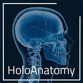
Case Western Reserve University School of Medicine developed the HoloAnatomy Software Suite in 2016 using Microsoft’s HoloLens 2 mixed reality headsets to visualize the body as a 3-dimensional hologram. The HoloAnatomy curriculum, the first third-party application for the Microsoft HoloLens device, launched in fall 2019, opening just a few months after the new Health Education Campus (HEC) of Case Western Reserve and Cleveland Clinic. When Case Western shifted classes online, the university shipped 97 HoloLens headsets to each of its anatomy students in order to hold a remote mixed reality class. You can view a pilot Hololens anatomy lesson here. Episode 42 of the All Access Med School Admissions podcast discusses the use of HoloAnatomy at Case Western Reserve.
Since then, TCU School of Medicine and Northwestern University Feinberg School of Medicine have followed suit. TCU integrated the technology at the beginning of each week of their course to introduce students to anatomy before the week’s “boot camp.” Later, they attend the cadaver lab to physically examine those structures. Northwestern plans to introduce it to first and second-year medical students in their Phase 1 Module. It will not be replacing traditional dissection but to augment the curriculum. Many schools are moving towards a prosection-based learning model rather than cadaver dissections. Their revamped anatomy curriculum includes rotation through different clinical cases with integrated medical imaging, histopathology content, and anatomy of prosecuted specimens augmented by the 3d HoloLens visualization. Students are still able to sign up for optional dissection sessions offered with each learning module. Other schools, such as the University of Central Florida College of Medicine, and California Health Science University’s College of Osteopathic Medicine, are evaluating how they might integrate the technology into their curriculum. The Journal of Ultrasound Medicine. November 2021. 40(11): 2459-2465)
The A.T. Still University’s Kirksville College of Osteopathic Medicine has integrated ultrasound into its first and second-year curriculum since 2011, and they started by integrating it into the Gross Anatomy I and II as a tool to visualize “living anatomy”. A survey in 2014 of the 134 U.S. MD granting schools demonstrated that 62.2% reported ultrasound training integrated into their UME curriculum, most commonly in the third year. A survey in 2021 demonstrated that 72.6% indicated they had an ultrasound curriculum, with 73.8% integrating it into the basic science courses (Nicholas E, et. al. The Current Status of Ultrasound Education in United States Medical Schools. The Dr. Kiran C. Patel College of Osteopathic Medicine at Nova Southeastern University is using BodyViz 3D anatomy dissection software with an option to work on cadavers.
Some students who really enjoy anatomy or who are pursuing surgical training may want more experience in anatomical study or dissections. Some medical schools have programs that allow students to participate in the protection preparation or to serve as teaching assistants for the anatomy curriculum.
Cleveland Clinic Lerner College of Medicine has an anatomy course for Physician Assistants and CCLCM medical students are able to participate as teaching assistants. In this role, they prepare the cadaver dissections and instruct the PA students. During the second year, students can assist the fellows and residents as they prepare the prosecution for the first-year students and for their review of anatomy in the second year. There are also several clinical/surgical anatomy electives that students can participate in.
One-year anatomy fellowships are common in osteopathic medical programs, some granting a Master’s degree including Rocky Vista School of Osteopathic Medicine, ACOM, Kansas City University College of Osteopathic Medicine, and Debusk College of Osteopathic Medicine.
EVMS offers a Master of Science Degree completed in 3 semesters in Contemporary Human Anatomy where there is an opportunity to serve as a TA for medical school students.
For those looking for a post bacc or summer experience I came upon this program (unpaid) that allows for anatomy dissection and research without any prior experience Seattle Science Foundation Clinical Anatomy Research Fellowship.